
Bone is a multiscale hierarchical structure composed of collagen, water, and mineral nanoparticles. Arrangements of these components into different functional units creates a light but tough structure that is multi-functional and able to adapt to diverse mechanical environments. Bone mass or bone mineral density is the parameter that is most commonly used to determine bone deterioration with age and to predict bone susceptibility to fracture. However, recent research clearly shows that reduced bone mineral density is not the sole factor in increased fracture risk. A complete understanding of bone fracture can be achieved by studying active fracture/ bone toughening mechanisms at different levels of its structure. In MCMB LAB, we focus on how bone structure and material properties at different scale influence the behavior of fractures. We employ the computational tools (e.g., the finite element method (FEM)) and mathematical techniques (e.g., the solution for edge dislocations, in terms of Green’s function), to study mechanics fracture of Haversian cortical bone. We evaluate our models by performing experimental tests (e.g., fracture test) and fractographical studies (e.g., scanning electron micrographs (SEM)).
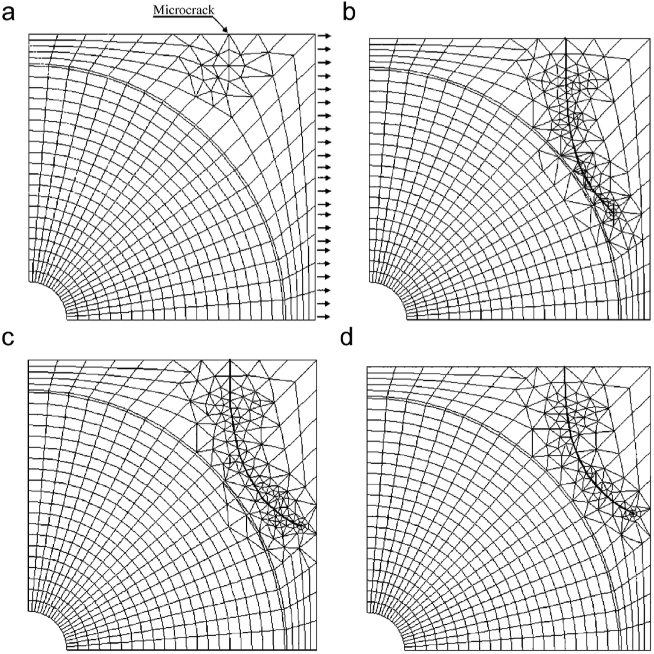 Microcrack propagation trajectory under tension: the propagation microcrack trajectory deviates as it approached the osteon. (a) Primarymicrocrack, and (b, c, d) propagation microcrack; (b) Eo = 10 GPa, Ei = 26 GPa, Ec = 6 GPa; (c) Eo = 19 GPa, Ei = 26 GPa, Ec = 6 GPa;(d) Eo = 19GPa, Ei = 15GPa, Ec = 24GPa.This work was done in collaboration with Dr. Arshi and Dr. Eslami (AUT).
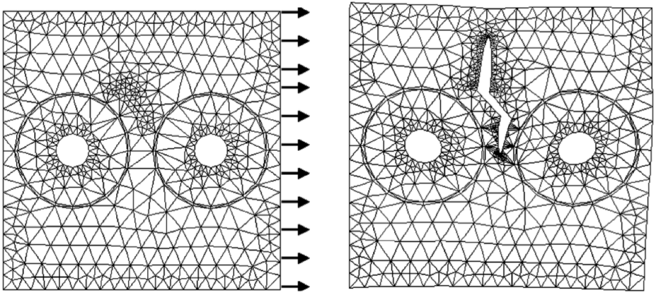 Microcrack propagation in between osteons during tensile loading.
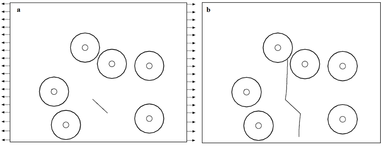 Internal microcrack interacting with multiple osteons, a) Primary microcrack, b) Propagation of microcrack. Crack comes to complete stop at the cement line region (at the boundary of osteon).
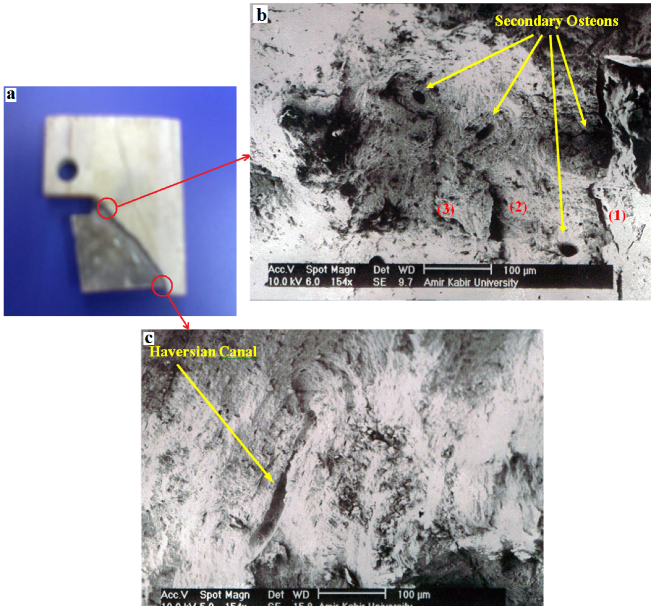 a) Fracture CT sample b) SEM of fracture surface at main crack initiation c) SEM of fracture surface at the main crack end. This work was done in collaboration with Dr. Arshi and Dr. Eslami (AUT).
Related Publications:
- Raeisi Najafi A., PourAkbar Saffar K., Arshi A. R., Mallakin E., Katouzian H.R., Eslami M. R., Moeinzadeh M. H.; “Propagation of Microcracks in Bovine Osteonal Cortical Bone”, Akademeia, vol. 1 (1): ea 103, 2011.
- Raeisi Najafi A., Arshi A. R., Kaveh PorAkbar Saffar, Eslami M. R., Fariborz S., Moeinzadeh M. H.; “A Fiber-Ceramic Matrix Composite Material Model for Osteonal Cortical Bone Micromechanics Fracture: General Solution of Microcracks Interaction” Journal of the Mechanical Behavior of Biomedical Materials, vol. 2, pp. 217-223, 2009
- Raeisi Najafi A., Mallakin E., PourAkbar Saffar K., Katouzian H.R., Arshi A.R., Eslami M.R.; “Fracture Test of Bovine Cortical Bone, Evaluation of Bone Toughness Properties and Consideration of Bone Microstructure Effect on Fracture Behavior” Amirkabir Journal of Science and Technology, vol. 68A, pp. 1-9, 2008.
- Raeisi Najafi A., Arshi A. R., Eslami M. R., Fariborz S., Moeinzadeh M. H.; “Micromechanics Fracture in Osteonal Cortical Bone: A Study of the Interactions between Microcrack Propagation, Microstructure and the Material Properties” Journal of Biomechanics, vol 40, pp. 2788-2795, 2007.
- Raeisi Najafi A., Arshi A. R., Eslami M. R., Fariborz S., Moeinzadeh M. H.; “Haversian Cortical Bone Model with Many Radial Microcracks: An Elastic Analytic Solution” Medical Engineering and Physics, vol. 29, pp. 708–717, 2007.
- Raeisi Najafi A., Arshi A. R., Eslami M. R., Fariborz S.; Moeinzadeh M. H.; “Micromechanics Fracture of Haversian Cortical Bone: An Analytical Approach” Transaction of ISME, vol 7, No 1, pp. 17-30, 2006.